|
Aubrey Funke, the Assistant Director of the Imaging and Histology Core Facility (IHCF), welcomed the iCREATE students to the IHCF to learn how to use an extremely powerful (and extremely expensive) microscope! The Scanning Electron Microscope (SEM) can magnify 1,000,000 times - which is 1,000 better than a light microscope, which is already 1,000 time better than our eyes! The Imaging and Histology Core Facility provides imaging services to faculty, undergraduate and graduate students, along with industry partners. They provide services in light microscopy, electron microscopy, and histology (microscopy of tissues). 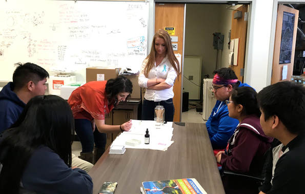 Alyssa Talbert, shows the students how to stain their samples while Aubrey looks on Graduate Student Alyssa Talbert creates scaffolds for wound healing with elastin, collagen and water in her graduate work. But here, she is illustrating how to collect and stain cheek samples for the students to view under the light microscope. 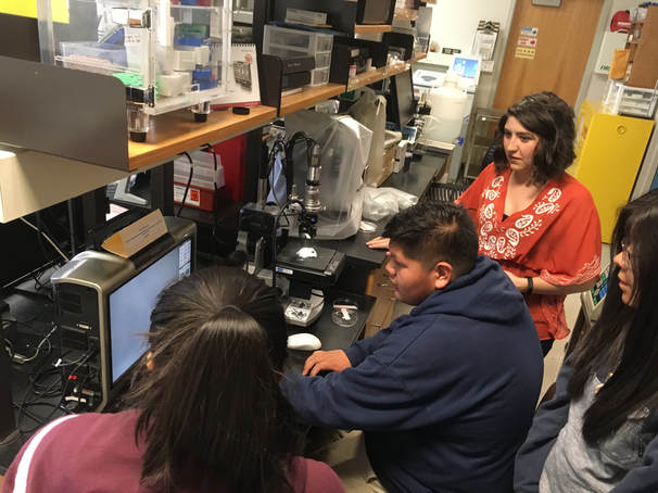 The students each practice using the digital light microscope Alyssa and Aubrey then take the students to compare the digital light microscope images with the detail they can see with the SEM. Because electrons have a very small wavelength, much smaller than the wavelength of light, the resolution using the SEM is much greater than that of light microscopes. 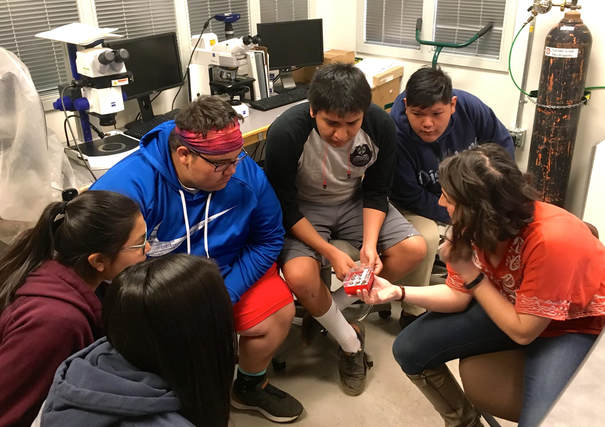 Alyssa shows the students how the samples get mounted on the stub for the SEM Aubrey and Alyssa shared a variety of SEM images with the students, from insects (see past Ugly Bug Contest winners below), to fungi, Alyssa's scaffolds, and much more!  Ugly Bug Contest poster from the 1996 Flagstaff Festival of Science The students spent approximately 8 hours at the IHCF learning how to use the SEM. They took copious notes they then followed explicitly, with Aubrey observing them, and they were able to run the SEM themselves on their last day of the training! 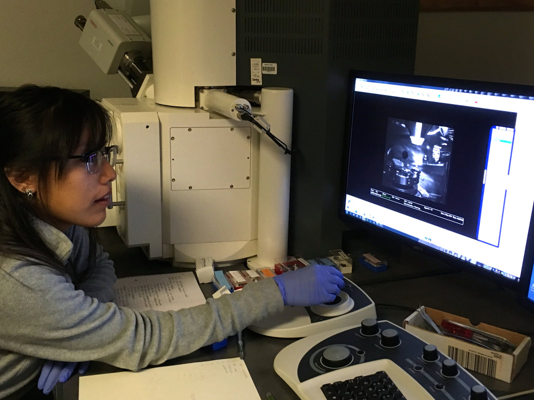 Mashayla Tso carefully lowers the stage on the SEM so she can observe her sample Thank you to both Alyssa and Aubrey for all the time and patient effort they put into instructing the students on the SEM. We weren't surprised to learn that Aubrey won the "Outstanding Staff Member" for NAU's Biology Department at their award ceremony on Wednesday, May 2nd. Congratulations to Aubrey! 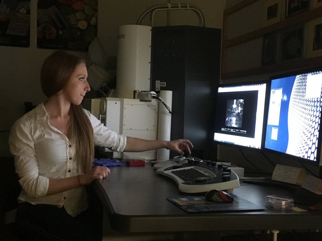 Aubrey focusing in on a sample on the SEM
3 Comments
Hi,thanks a lot for this information. I loves to read this blog. Keep it up Leave a Reply. |
